Common Eye Problems and What They Look Like LSC Eye
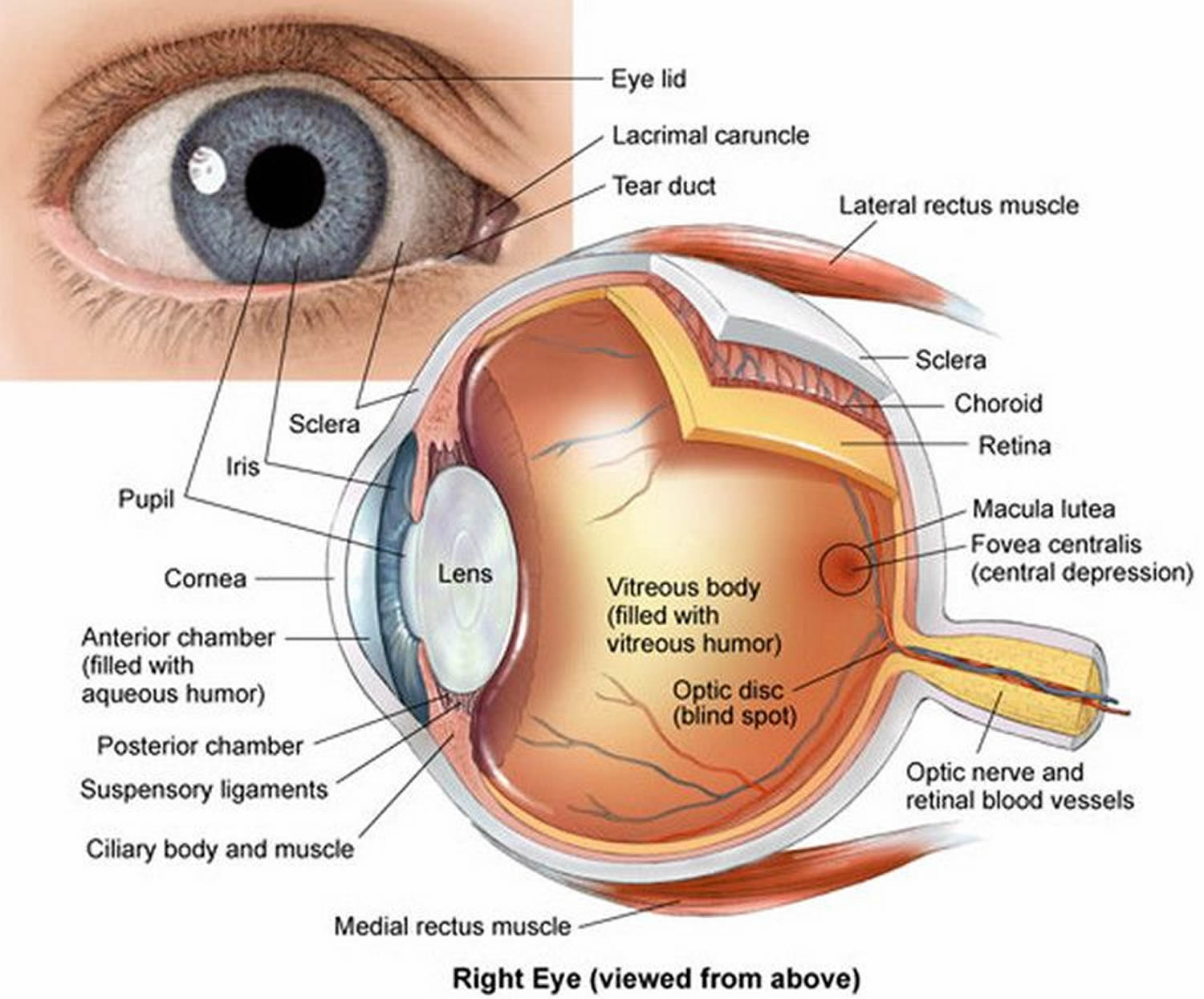
View All. Cornea. Pupil. Iris. Crystalline Lens. Aqueous Humor. The human eye is an organ that detects light and sends signals along the optic nerve to the brain. Perhaps one of the most complex organs of the body, the eye is made up of several parts—and each individual part contributes to your ability to see.
Regularity pause Linguistics lens eye Match Ambient Resume
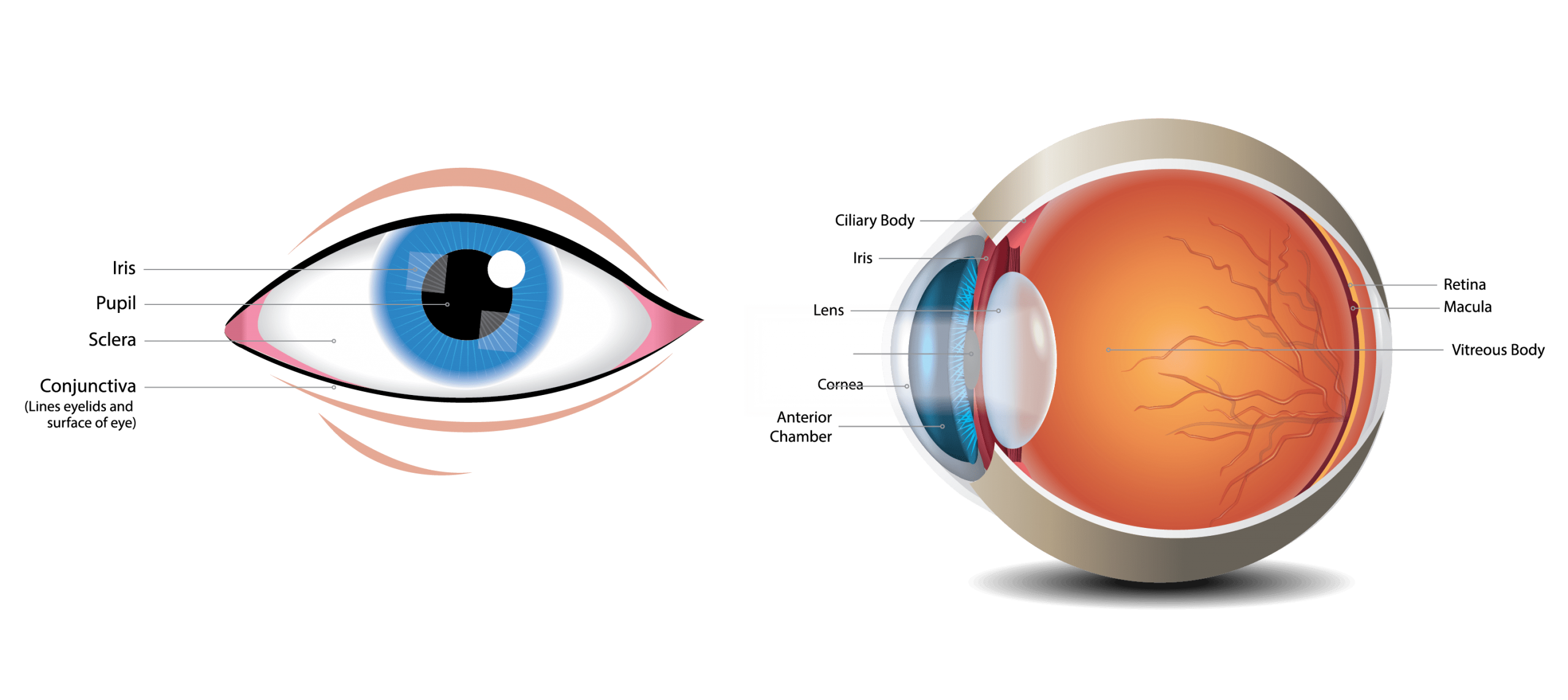
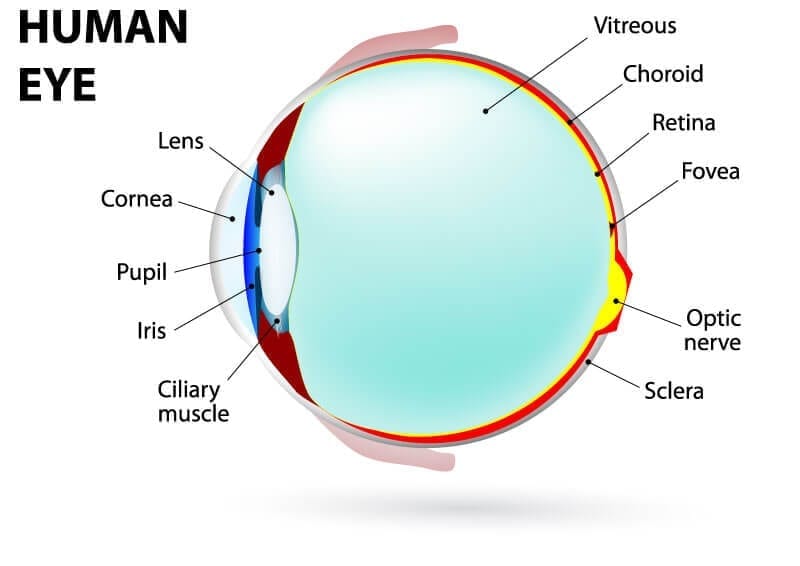
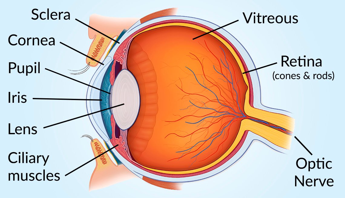
The main parts of the human eye are the cornea, iris, pupil, aqueous humor, lens, vitreous humor, retina, and optic nerve. Light enters the eye by passing through the transparent cornea and aqueous humor. The iris controls the size of the pupil, which is the opening that allows light to enter the lens. Light is focused by the lens and goes.
Inflammatory Arthritis and Eye Health Prevention, Symptoms, Treatment
Picture of Eye Anatomy Detail. The eye is our organ of sight. The eye has a number of components which include but are not limited to the cornea, iris, pupil, lens, retina, macula, optic nerve, choroid and vitreous. Cornea: clear front window of the eye that transmits and focuses light into the eye. Iris: colored part of the eye that helps.
Internal Parts and Functions of the Eye HubPages
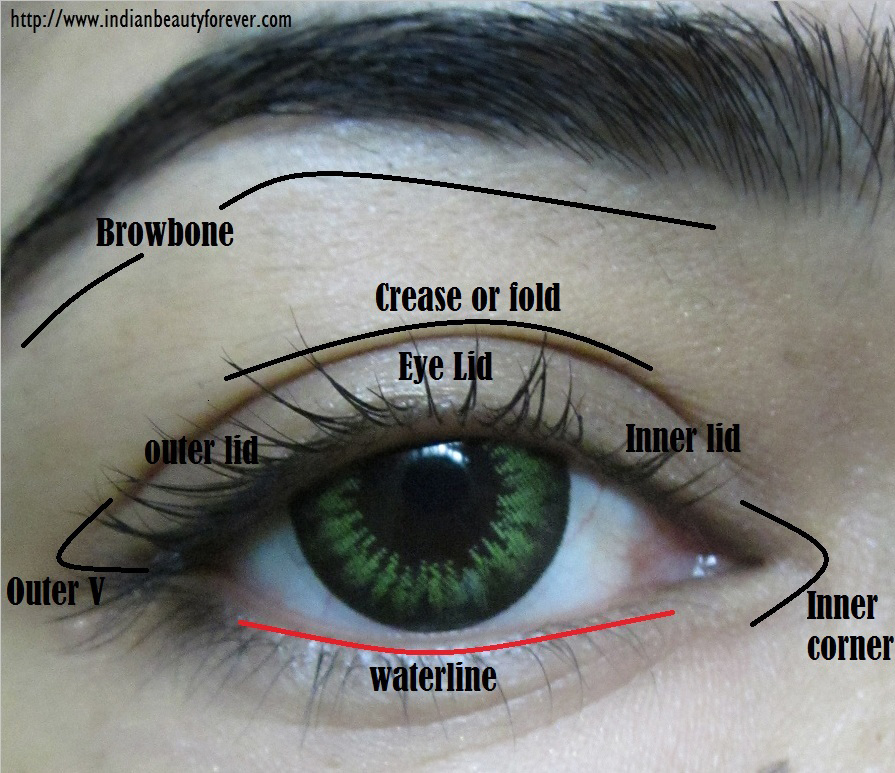
The front part (what you see in the mirror) includes: Iris: the colored part. Cornea: a clear dome over the iris. Pupil: the black circular opening in the iris that lets light in. Sclera: the.

OUR EYES WORK LIKE CAMERA’S! Discovery Eye Foundation
Parts of the eye, labeled vector illustration diagram Parts of the eye, labeled vector illustration diagram. Educational beauty and nursing information.. vision, catheract, ostegmatism, laser eye surgery. 3D illustration, 3D render. human eye anatomy stock pictures, royalty-free photos & images. Realistic human eye hologram, cross sectional.
Human Eye Anatomy Parts of the Eye and Structure of the Human Eye
human eye, in humans, specialized sense organ capable of receiving visual images, which are then carried to the brain.. Anatomy of the visual apparatus Structures auxiliary to the eye The orbit. The eye is protected from mechanical injury by being enclosed in a socket, or orbit, which is made up of portions of several of the bones of the skull to form a four-sided pyramid, the apex of which.
Ashes to Ashes March 2013
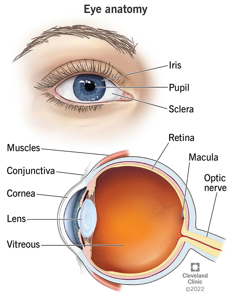
The diagram below shows the parts of the human eye that work together to enable us to see. These include the lens, retina and the optic nerve. Other parts of the eye include the aqueous humour, which is a liquid that sits in a chamber behind the cornea. It keeps the eye nourished and helps it maintain an optimum pressure so that the eyeball.

Share
The iris (colored part) of the eye functions like the diaphragm of a camera, controlling the amount of light reaching the retina by automatically adjusting the size of the pupil (aperture). The eye's crystalline lens is located directly behind the pupil and further focuses light rays. Through a process called accommodation, this lens.
/GettyImages-695204442-b9320f82932c49bcac765167b95f4af6.jpg)
Structure and Function of the Human Eye
Iris - Coloured circle around the pupil. It controls the size of the pupil. Pupil - Black part of the eye. This is an opening that lets light in. Retina - Light-sensitive layer at the back of the.
Understanding the Different Parts of Your Eye All About Eyes
The optic nerve carries signals of light, dark, and colors to a part of the brain called the visual cortex, which assembles the signals into images and produces vision. Posterior chamber. The back part of the eye's interior. Pupil. The opening in the middle of the iris through which light passes to the back of the eye. Retina.

Can We Grow New Eyes?
The structures and functions of the eyes are complex. Each eye constantly adjusts the amount of light it lets in, focuses on objects near and far, and produces continuous images that are instantly transmitted to the brain. The orbit is the bony cavity that contains the eyeball, muscles, nerves, and blood vessels, as well as the structures that.

Which Parts of the Eyes Are Associated with Which Eye Diseases? Natural Eye Care Blog News
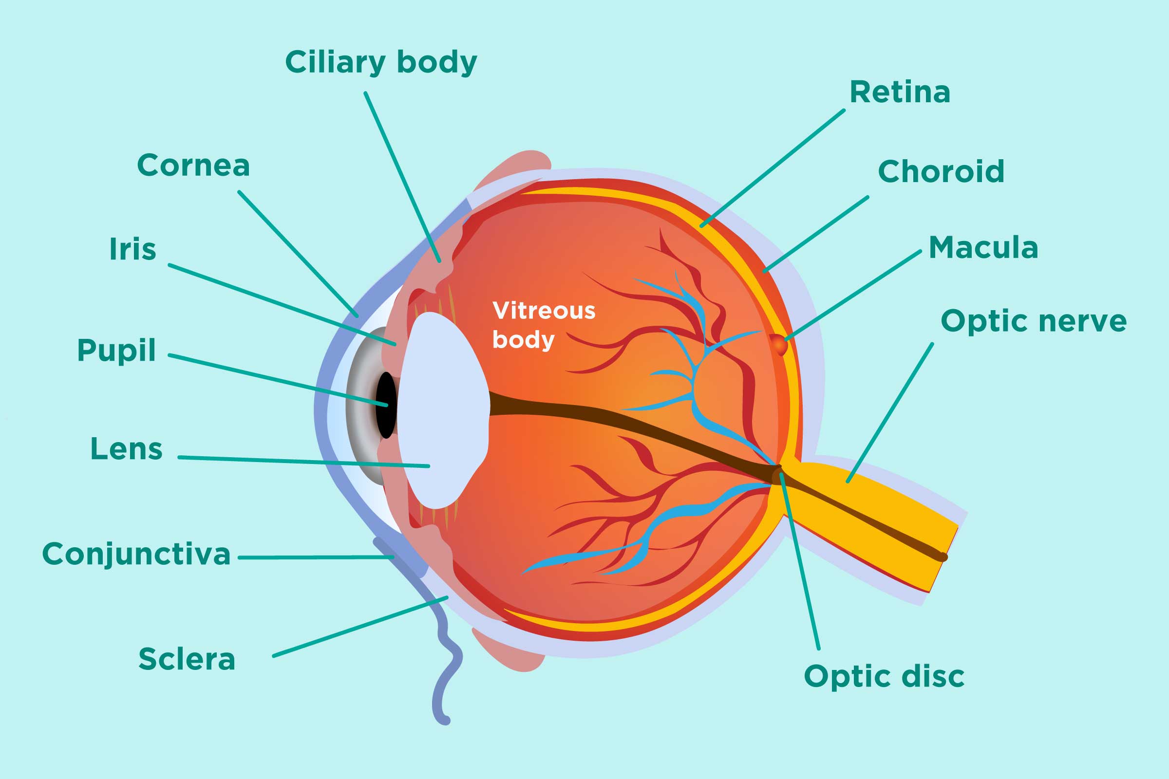
Ciliary body: the part of the eye that connects the choroid to the iris. Retina: a light sensitive layer that lines the interior of the eye. It is composed of light sensitive cells known as rods and cones. The human eye contains about 125 million rods, which are necessary for seeing in dim light. Cones, on the other hand, function best in.
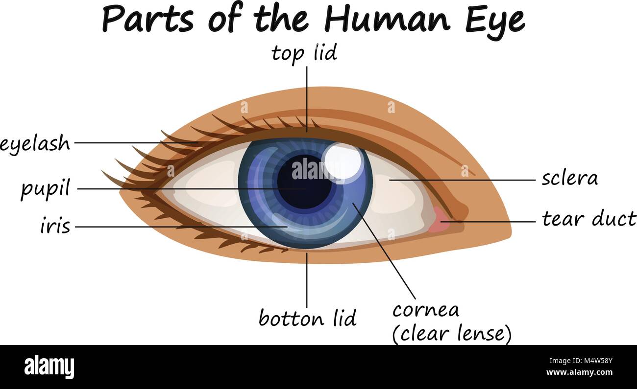
Diagram showing parts of human eye illustration Stock Vector Image & Art Alamy
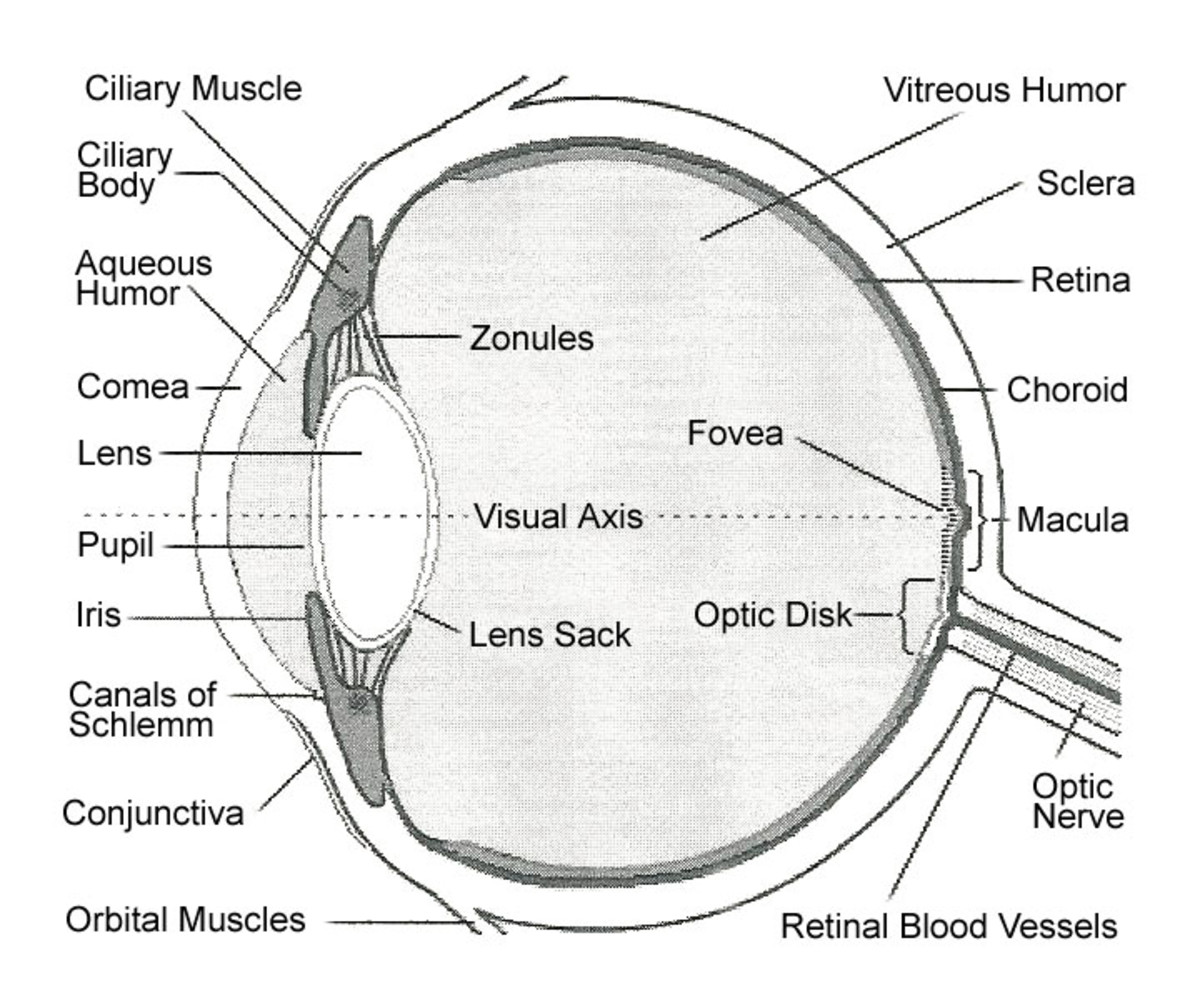
The light passing through cornea, pupil, and lens gets focused on the retinal membrane. In addition to tissue components, retina is made up of two types of cells: rod cells and cone cells. The.

Picture Of Eye Parts YouTube
However, to prevent damage to the cornea and other parts of the eye, you should wear sunglasses. Chronic exposure to UV light may lead to inflammation and other complications, including cancer. 4. 6. Uvea. The uvea is the eye's middle layer. It is located underneath the white part of the eye (the sclera) and is composed of three parts: The iris.

Human Eye Anatomy Parts of the Eye and Structure of the Human Eye
When the eye is directed at an object, the part of the image that is focused on the fovea is the image most accurately registered by the brain. • Iris: regulates the amount of light that enters your eye. It forms the coloured, visible part of your eye in front of the lens. Light enters through a central opening called the pupil.
Vision and Eye Diagram How We See
Behind the anterior chamber is the eye's iris (the colored part of the eye) and the dark hole in the middle called the pupil. Muscles in the iris dilate (widen) or constrict (narrow) the pupil to control the amount of light reaching the back of the eye. Directly behind the pupil sits the lens. The lens focuses light toward the back of the eye.